
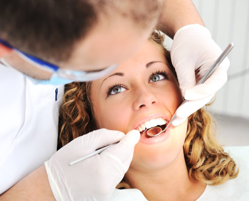
This page brings together the 75 questions international patients ask most often about Oral and Maxillofacial Surgery for International Patients in India. Each answer explains the clinical reasoning and ends with a Patient Decision Check you can take to your consultation.
Referral, Diagnosis and Pre-Travel Evaluation: Oral and Maxillofacial Surgery for International Patients in India
01. When should an international patient consider oral and maxillofacial surgery in India?
Consider planned treatment abroad when a defined jaw, facial-bone, temporomandibular-joint or reconstructive condition requires specialist expertise, imaging-based planning, or a multidisciplinary team not easily accessible locally. Examples include severe jaw discrepancies, ankylosed jaw joints, complex facial fractures after initial stabilization, benign jaw tumours and reconstruction after cancer. An overseas consultation cannot replace emergency treatment for airway compromise, active bleeding or rapidly spreading infection. Selection should weigh expected functional benefit against travel, the need for orthodontics, staged reconstruction and postoperative monitoring. A technically feasible operation is not automatically the right operation for a patient who cannot arrange reliable follow-up at home.
02. What conditions are treated by an oral and maxillofacial surgeon rather than a general dentist or plastic surgeon?
Oral and maxillofacial surgeons treat conditions involving the jaws, facial skeleton, oral tissues and related functions, often combining dental occlusion with reconstructive principles. Their scope may include orthognathic surgery, TMJ reconstruction, complex facial fractures, jaw cysts, benign and selected malignant tumours, salivary problems and skeletal deformities. General dentists commonly handle routine tooth care, while plastic, ENT, head-and-neck and neurosurgical teams may participate in overlapping complex cases. The decisive issue is not the professional title alone but documented training and current experience with the precise operation. Many advanced cases need a combined surgeon, orthodontist, radiologist and anaesthesia plan.
03. Which scans, photographs and reports should I send before requesting a maxillofacial opinion from India?
Send a symptom chronology, previous surgical records, current medicines, photographs showing facial symmetry and bite, dental records and the original imaging files rather than only screenshot reports. Panoramic radiography is useful for broad dental-jaw screening; cone-beam CT or medical CT can define skeletal anatomy; MRI is particularly valuable for soft tissues and the TMJ disc. Facial trauma, tumours and suspected cancer may require contrast-enhanced imaging or dedicated staging. Include orthodontic records and digital dental models for planned jaw correction. The Indian team should explain which images are sufficient for preliminary planning and which will need repeating after clinical examination.
04. Can an Indian surgeon confirm my diagnosis and provide a fixed treatment plan through a video consultation?
A remote consultation can review reported symptoms, compare imaging, identify likely surgical pathways and provide an initial estimate, but cannot fully assess bite contacts, palpation, jaw function, airway or neurological deficits. Diagnoses such as TMJ pain, facial asymmetry or chronic swelling may have more than one cause. Complex reconstructions often change when updated CT scans, examination and dental assessments become available. Written recommendations should therefore distinguish a provisional plan from the final consented operation. Avoid paying nonrefundable fees on the assumption that an online assessment establishes eligibility, operative time or the exact implant inventory. The final operative plan should therefore remain conditional until the in-person findings are documented.
05. How do surgeons determine whether facial asymmetry is caused by the jaws, teeth, muscles or a TMJ disorder?
Assessment compares the dental midlines, occlusal plane, facial thirds, chin position, mandibular movements and joint symptoms. Dental compensation can conceal skeletal asymmetry, while unilateral condylar overgrowth or resorption can progressively change facial proportions. A panoramic image, cephalometric analysis and CBCT or CT may be useful; MRI or functional studies are added for suspected active joint disease. Treating only the chin may improve appearance without correcting the bite or continuing abnormal jaw growth. Conversely, not every visible asymmetry requires major jaw surgery. The plan must identify whether the underlying skeletal process is stable before moving bones or placing implants.
06. Which facial or jaw symptoms mean I should seek emergency care locally rather than travel to India?
Difficulty breathing, inability to swallow saliva, rapidly spreading facial or neck swelling, uncontrolled bleeding, a new major neurological deficit or unstable facial trauma requires immediate local assessment. Fever with trismus, floor-of-mouth swelling or voice change can indicate a deep-space infection threatening the airway. Visual loss or severe eye pain after an orbital injury is also urgent. Delaying treatment to catch an international flight can worsen outcomes, and commercial air travel may be inappropriate during acute infection, bleeding or unstable fractures. Medical tourism is better suited to planned reconstruction after the immediate danger is controlled. Airway deterioration can evolve quickly, making delay especially dangerous in deep neck infections.
07. What is the difference between CBCT, conventional CT and MRI for maxillofacial surgery planning?
Cone-beam CT shows dental roots and facial bones at useful detail, often with lower radiation burden than some medical CT protocols, but its soft-tissue assessment is limited. Medical CT is more suitable for complex fractures, extensive tumours and cases requiring contrast-enhanced evaluation of soft tissue or vessels. MRI helps identify TMJ discs, joint effusion, muscles and selected soft-tissue lesions without ionizing radiation. The correct scan depends on the suspected disease, not a rule that every patient requires three-dimensional imaging. Images should be reviewed together with the examination and, when relevant, histopathology; a high-resolution scan cannot determine whether a lesion is benign by appearance alone.
08. How should patients with diabetes, heart disease or blood-thinner treatment prepare for major jaw surgery?
Major maxillofacial procedures may involve prolonged general anaesthesia, bleeding risk, restricted oral intake and infection concerns. Preoperative evaluation should identify poorly controlled diabetes, anaemia, anticoagulant or antiplatelet use, cardiovascular limitations, kidney dysfunction, malnutrition and obstructive sleep apnoea. Patients must not stop heart or blood-thinning medicines independently; the prescribing specialist and anaesthetist should agree on any changes. Some procedures require dental infection clearance or nutritional support before surgery. Complex conditions can justify inpatient monitoring or staged treatment instead of an outpatient package. A low quoted price cannot compensate for inadequate perioperative medicine support. Smoking and alcohol intake should also be discussed because they can impair healing.
09. Can children and adolescents travel to India for jaw deformity or craniofacial surgery?
Selected children with congenital jaw differences, post-traumatic growth disturbances, cleft-related deformities or TMJ ankylosis may benefit from specialist care. Timing differs greatly by diagnosis. Some operations protect breathing, feeding or normal development and should not be delayed; elective orthognathic corrections often wait until facial growth is sufficiently mature. Growth modification, distraction osteogenesis and staged reconstruction may be considered in selected cases, with continuing review as the child grows. Parents should understand that one operation may not end treatment. Paediatric anaesthesia, growth-sensitive planning and coordination with local orthodontists or speech therapists are essential for overseas families.
10. How can I compare two Indian surgeons recommending different maxillofacial operations?
Begin by comparing the exact diagnosis and the problem each plan is designed to solve: bite correction, jaw opening, airway improvement, tumour control or facial reconstruction. Different techniques may both be defensible when anatomy and priorities vary. Request the planned bone movements or resection boundaries, imaging rationale, surgeonspecific experience, expected functional limitations, major risks and alternatives. For orthognathic cases, the associated orthodontic strategy is fundamental; for tumour surgery, histology and margins matter more than an attractive threedimensional simulation. An independent multidisciplinary opinion can reveal whether a disagreement reflects genuine uncertainty or incomplete records. For cancer-related operations, confirm whether the proposed surgeon participates in a formal tumour board.
Corrective Jaw Surgery and Early TMJ Decisions: Oral and Maxillofacial Surgery for International Patients in India
11. What is orthognathic surgery, and when are braces alone insufficient to correct a bite?
Orthognathic surgery repositions the upper jaw, lower jaw or both to address skeletal discrepancies affecting chewing, speech, facial balance or sometimes breathing. Braces move teeth within bone but cannot reliably correct a large mismatch between the jaw bases once growth is complete. A severe underbite, overbite, open bite or facial cant may therefore require coordinated orthodontic and surgical treatment. An orthodontist must assess whether apparently crooked teeth are compensating for the underlying deformity. Surgery is not appropriate solely because someone dislikes their profile; expected functional improvements, nerve risks and long-term stability deserve equal weight. The proposed occlusion must also remain maintainable after braces are removed and retained.
12. How do Le Fort I, bilateral sagittal split osteotomy and genioplasty differ?
A Le Fort I osteotomy repositions the upper jaw; a bilateral sagittal split osteotomy changes lower-jaw position; genioplasty reshapes or repositions the chin without moving the bite-bearing jaw segments. These procedures can be combined, but each addresses different anatomy. Upper-jaw movement can change nasal appearance, incisor show and bite alignment, while lower-jaw surgery carries particular sensory-nerve risks. A chin-only operation may correct projection without improving a skeletal malocclusion. Surgeons use clinical examination, dental models and threedimensional planning to determine movements and safe fixation. The least extensive operation that meets the patient's functional goals is generally preferable.
13. Is double-jaw surgery more effective than single-jaw surgery for severe facial deformity?
Double-jaw surgery allows coordinated repositioning of the upper and lower jaws and may better correct complex open bites, facial cants, occlusal-plane problems or large discrepancies. However, it usually involves longer surgery and may increase swelling, cost and the number of structures at risk. Single-jaw surgery can be entirely appropriate when the other jaw is well positioned. The choice should emerge from cephalometric analysis, facial examination, stable joint status and orthodontic planning, not from a promise that more extensive surgery always gives a better result. Threedimensional simulations help explain the plan but cannot guarantee symmetry or exact final soft-tissue appearance.
14. How long must I wear braces before and after corrective jaw surgery if I live abroad?
Traditional orthognathic treatment commonly involves a pre-surgical orthodontic phase to align and decompensate teeth, followed by surgery and further orthodontics to refine the bite. The total course can extend over many months or longer, depending on the discrepancy and tooth movement required. Some selected patients may be candidates for a surgery-first protocol, but this demands careful screening and reliable orthodontic follow-up. International patients who expect one short surgical trip must identify an orthodontist at home willing to work to the Indian surgeon's plan. Interrupting braces adjustments can compromise final occlusion and stability. Request a strategy for missed appointments if travel restrictions or illness disrupt the schedule.
15. What is virtual surgical planning, and does 3D printing make jaw correction safer?
Virtual planning combines CT or CBCT imaging with digital dental models to simulate bone movements, design splints and sometimes guide fabrication of patient-specific plates or cutting guides. It can improve communication and help anticipate complex asymmetry or anatomy. Yet virtual accuracy depends on correct bite registration, stable joints and the surgeon's ability to execute the plan safely. Printed models and custom hardware add cost and may need manufacturing lead time; they do not eliminate bleeding, nerve injury or relapse. Conventional planning remains effective for many routine cases. A claim that a custom plate guarantees a perfect face or zero revision risk is not credible.
16. Can corrective jaw surgery improve obstructive sleep apnoea, and who qualifies?
Maxillomandibular advancement can enlarge the upper-airway framework in selected patients with obstructive sleep apnoea, particularly when craniofacial anatomy contributes to airway obstruction. It is not a general replacement for evidence-based treatment such as positive-airway-pressure therapy. Sleep-study severity, symptoms, body habitus, dental condition, airway examination and tolerance of other treatments influence selection. Advancing the jaws changes facial appearance and carries the risks of major osteotomy. Postoperative sleep testing is needed because improvement is not identical to a guaranteed cure. An international patient must arrange ongoing sleep-medicine care, especially if residual disease or device use remains necessary.
17. Can orthognathic surgery treat an open bite, gummy smile or facial asymmetry without relapse?
Jaw surgery can address selected skeletal open bites, excessive vertical maxillary growth and facial cants when orthodontics alone is inadequate. Stability depends on the type and direction of movement, quality of fixation, condylar position, muscle adaptation and orthodontic control. Some movements have higher relapse concerns, especially in patients with active condylar resorption or untreated joint disease. Soft tissues may not follow skeletal movements exactly, so the final smile cannot be guaranteed from a screen rendering. Patients need a long-term retention strategy and an honest discussion of residual asymmetry or secondary refinements. Long-term photographs and bite records are more informative than an immediately postoperative simulation.
18. Is surgery-first orthognathic treatment suitable for patients who want one short visit to India?
Surgery-first protocols place the jaws before extensive preoperative braces and may be feasible for carefully selected patients with predictable dental relationships. They are not a shortcut that removes the need for orthodontics. Postoperative tooth movements can be demanding and depend on frequent appointments, bite adjustment and communication between the surgical and orthodontic teams. Severe crowding, unstable occlusion, joint problems or complex asymmetry may favor the traditional sequence. Travelling home without an experienced orthodontist able to finish the process exposes patients to bite deterioration and avoidable revision. Programmes should describe selection criteria and share a realistic timeline rather than advertise instant completion.
19. What nerve injuries, facial numbness and bite changes are possible after double-jaw surgery?
Lower-jaw osteotomies run near the inferior alveolar nerve, which supplies sensation to the lower lip and chin. Temporary altered sensation is common, while persistent numbness or abnormal tingling can occur. Upper-jaw surgery may affect sensation in the upper lip, cheeks or teeth and can change nasal shape or breathing symptoms. Bite changes may arise from swelling, muscle adaptation, fixation or condylar positioning. Nerve recovery can take months and is not guaranteed. Surgeons should also discuss infection, hardware problems and the possibility of additional orthodontic treatment. An international patient should not infer minimal risk from hidden intraoral incisions.
20. How is jaw fixation performed, and will titanium plates or screws need removal later?
Most modern corrective jaw procedures use small titanium plates and screws to stabilize repositioned bone while it heals. Elastics or guiding splints may help the bite settle, but routine prolonged wiring shut is not always necessary. Plate design varies with movement and bone quality. Hardware often remains in place permanently unless infection, exposure, discomfort or another problem requires removal. An implant label alone does not establish quality; positioning, fixation stability and infection prevention are essential. Patients should receive details of the actual material and postoperative instructions, including what to do if a screw loosens or a wound starts draining.
21. What does TMJ pain mean, and when should a patient be investigated for an internal joint disorder?
Temporomandibular symptoms include jaw pain, clicking, locking, headache, muscle tenderness and difficulty chewing. Not every symptom arises from structural joint damage; teeth clenching, muscle pain and other pain conditions may contribute. Examination assesses opening distance, deviation, bite, tenderness and joint sounds. MRI can identify disc displacement or inflammation when findings would affect treatment, while CT evaluates bony degeneration or ankylosis. Most patients need an initial non-surgical approach rather than immediate joint replacement. Previous failed treatments and the degree of functional disability matter in deciding whether further investigation is worthwhile. An assessment for dental disease and associated headache conditions may prevent unnecessary operations.
22. When is TMJ arthrocentesis preferable to arthroscopy or open joint surgery?
Arthrocentesis uses joint lavage and sometimes injection to address selected inflammatory or painful internalderangement problems after conservative treatment. Arthroscopy adds direct visualization and the ability to perform limited procedures inside the joint. Open surgery is reserved for situations needing broader access, such as severe structural pathology or failed less-invasive management. None of these methods is a universal cure for muscular jaw pain or centrally mediated chronic pain. Outcomes depend on diagnosis, disease stage, previous treatment and physiotherapy participation. Patients travelling from abroad should compare the burden of surgery with the benefit likely from a carefully supervised local conservative programme.
23. When is total temporomandibular joint replacement recommended in India?
Total TMJ replacement is considered for selected patients with end-stage joint destruction, ankylosis, severe osteoarthritis, condylar loss or multiple failed reconstructive operations when function remains substantially impaired. It replaces both the mandibular and temporal joint components using a stock or patient-specific prosthetic system. Most patients should first undergo appropriate diagnosis and consideration of less invasive choices. Joint replacement may improve opening, chewing and pain, but does not guarantee normal motion or complete pain elimination. Implant availability, allergy history, previous infection, opposing joint condition and expected rehabilitation all influence selection. Patients require continued surveillance of a permanent device.
24. What is the difference between stock and patient-specific custom TMJ prostheses?
Stock TMJ systems use premanufactured component sizes adapted to a patient's anatomy, whereas custom devices are designed from imaging to fit the individual skull and jaw. Custom components may be particularly valuable for severe deformity, previous surgery or asymmetrical bone loss, but may cost more and require additional manufacturing time. Both depend on secure fixation, accurate occlusion, clean operative fields and careful postoperative physiotherapy. A custom-made device does not automatically have better longevity or freedom from infection. For patients travelling internationally, revision availability and future access to compatible replacement components matter almost as much as the initial operation.
25. How is TMJ ankylosis treated when the mouth can barely open?
TMJ ankylosis involves fibrous or bony restriction of joint movement and may follow trauma, infection or inflammatory disease. Severe cases can affect nutrition, oral hygiene, speech and airway access. Treatment may include release of the fused area, interpositional tissue, joint reconstruction or total replacement depending on age, deformity and recurrence history. Children may have associated mandibular growth disturbance, making planning especially complex. An anaesthetist experienced with difficult airways is essential because conventional mouth opening can be impossible. Postoperative exercises are crucial, and recurrence remains possible if rehabilitation is interrupted. Overseas patients should arrange supervised physiotherapy after returning home.
TMJ Reconstruction, Facial Trauma and Jaw Lesions: Oral and Maxillofacial Surgery for International Patients in India
26. Can TMJ surgery correct chronic jaw locking without making facial pain worse?
The surgical choice depends on whether the locking reflects a displaced disc, adhesions, degenerative change, muscle spasm or another problem. Intermittent clicking without major functional limitation usually does not justify invasive surgery. Persistent closed lock with restricted opening may sometimes respond to appropriately selected minimally invasive treatment. Chronic pain may have muscular, neuropathic and psychological contributors that remain after joint mechanics improve. An operation directed only at the disc may not resolve all symptoms and carries risks of facialnerve irritation, scarring or persistent stiffness. A multidisciplinary pain and rehabilitation plan can be as important as the operation itself.
27. How do surgeons decide whether a painful TMJ needs disc repair, arthroplasty or complete replacement?
MRI findings, degree of bone destruction, mouth opening, bite changes, inflammatory disease, joint loading and prior operations guide selection. A relatively preserved joint with a repairable disc may permit disc procedures; severe erosion, ankylosis or failed reconstruction can favor replacement. The treatment must also assess the opposite joint and any progressive condylar resorption, especially before orthognathic surgery. Disc displacement visible on MRI does not automatically require repair because asymptomatic displacement occurs. Operative consent should compare expected gains in opening and function against risks of nerve weakness, prosthesis infection and persistent pain. Ask whether simultaneous orthodontic adjustment is needed when joint position changes the bite.
28. Can bilateral TMJ replacement be combined with corrective jaw surgery?
Some patients with advanced joint destruction and severe skeletal malocclusion need reconstruction of both jaw joints plus orthognathic movements. Combining procedures may restore vertical jaw position and improve the bite, but increases operative complexity, blood loss, planning demands and the need for custom devices. Accurate virtual planning must account for the condyles, occlusion, teeth and upper-jaw position. The team may recommend a staged approach when infection, unstable tissue or medical conditions make combined surgery unsafe. International patients must plan for postoperative physiotherapy, possible orthodontics and several months of monitoring rather than a brief cosmetic-style recovery.
29. What happens if a previous TMJ implant becomes infected, loose or mechanically damaged?
Prosthesis problems may present as new swelling, fever, pain, bite alteration, reduced opening or abnormal imaging; not all discomfort means device failure. The surgeon must investigate for infection, loosening, bone loss, wear or changes in the opposite joint. Evaluation may include CT, inflammatory tests and specialist microbiology. A true deep implant infection can require removal, controlled infection treatment and later reconstruction rather than simple oral antibiotics. Revision may be difficult because of scarring and proximity to facial nerves and important vessels. Patients should not travel internationally with suspected acute infection unless a specialist has stabilized the problem.
30. What should international patients know about rehabilitation after TMJ replacement or ankylosis release?
Early mobilisation and structured jaw-opening exercises help reduce postoperative stiffness, but timing and intensity vary by fixation method and tissue condition. Surgeons may prescribe controlled stretching, physiotherapy, bite monitoring, a soft diet and follow-up imaging. Overaggressive force can cause pain or damage, while prolonged inactivity risks restricted opening. The patient should know expected mouth-opening milestones and whether specialised devices or a home physiotherapist are required. Long-haul travel does not replace weeks of rehabilitation. Patients with bilateral reconstruction may also need nutritional counselling because chewing endurance returns gradually and prosthetic joints have activity limitations. Exercise recommendations should be adapted promptly if pain, clicking or opening deteriorates significantly.
31. Which facial fractures need urgent surgery, and which can be repaired after initial stabilization?
Facial fractures vary from isolated stable nasal or zygomatic injuries to complex panfacial trauma. Immediate threats include airway obstruction, heavy bleeding, eye injury, intracranial injury and open contaminated wounds. Once a trauma team controls these, definitive reconstruction may be timed according to swelling, associated injuries and fracture displacement. Delay can make displaced bones harder to mobilise, so patients should not postpone urgent fixation simply to seek a cheaper overseas option. Stable older fractures with residual deformity may be considered for planned corrective reconstruction. The initial evaluation often needs CT of facial bones and assessment of vision, dental occlusion and neurological function.
32. How is a fractured lower jaw repaired, and will I be able to eat normally afterwards?
Mandibular-fracture management depends on fracture location, displacement, teeth in the line of fracture, occlusion and patient condition. Some fractures are treated by guided immobilisation, while others need open reduction and internal fixation with plates or screws. The surgeon must restore the bite and protect the inferior alveolar nerve. Infection, malunion, nonunion, hardware exposure and persistent numbness are possible. A soft or modified diet is commonly necessary while bone heals, with progression based on examination and imaging. Patients with associated condylar fractures may require jaw-opening exercises rather than extended immobilisation. Long-term dental follow-up is important. Associated tooth injuries can change fixation choices and should be recorded before treatment.
33. When do orbital-floor or cheekbone fractures require specialist reconstruction?
A zygomatic or orbital-floor fracture may alter facial projection, eye position, eye movement or sensation below the eye. Urgent assessment is necessary for visual loss, severe globe injury, muscle entrapment with systemic symptoms or other ophthalmic emergencies. For stable injuries, decisions about repair reflect diplopia, enophthalmos, displacement and CT findings, balanced against swelling and surgical risks. Reconstruction may use titanium mesh or patient-specific implants. Not every small orbital-floor defect needs surgery. An experienced maxillofacial surgeon should coordinate with ophthalmology when eye function is affected. Overseas travel before ophthalmic clearance can delay essential care. New double vision or changes in pupil appearance deserve prompt ophthalmic evaluation.
34. Can old facial fractures and post-traumatic asymmetry be corrected months or years later?
Secondary reconstruction can address malunited fractures, facial flattening, orbital volume problems, bite deformity and asymmetrical bone contours. It may require osteotomies to mobilise healed bones, grafts, custom implants or combined orthodontic treatment. Results can be limited by scar tissue, nerve injury, missing bone and previous hardware. A realistic plan separates improvements in appearance from recovery of vision, sensation or chewing, which may not fully return. CT-based three-dimensional analysis is especially helpful for comparing the affected and unaffected sides. Complex secondary surgery can take longer than initial fixation and may require several stages. Preoperative photographs help establish which asymmetries were present before the injury.
35. How are complex panfacial fractures reconstructed when several facial bones are damaged?
Panfacial injuries may involve the mandible, maxilla, zygomas, nasal-orbital-ethmoid region and orbital walls. Treatment typically prioritises a secure airway, associated brain and eye injuries, and restoration of facial height, width, projection and dental occlusion. Surgeons may use sequential fixation, existing teeth, splints and three-dimensional CT planning to recreate lost landmarks. Bone grafting or patient-specific implants are sometimes necessary. The risk of infection, malocclusion, diplopia and contour asymmetry rises with injury complexity. Patients arriving from another country should bring all initial trauma scans and operative notes so the reconstruction team understands what has already been repaired.
36. Can displaced cheekbones or facial bones be repaired without visible scars?
Some zygomatic, orbital or maxillary fractures can be accessed through incisions inside the mouth, along the eyelid or in inconspicuous facial creases. The approach depends on fracture extent and the need to see and stabilise specific bone segments. Avoiding an external incision should not compromise eye safety, nerve protection or stable fixation. Even well-placed incisions can leave scars or temporary facial nerve weakness. Highly displaced or secondary fractures may require wider access, grafting or custom plates. Patients should evaluate the expected functional repair and cosmetic trade-offs together, not select surgery based solely on a promise of scarless treatment.
37. What are the options for reconstruction after a high-energy facial injury with missing bone or soft tissue?
Severe injuries may leave defects in the jaw, cheek, orbit or mouth and require coordinated bone and soft-tissue reconstruction. Options include local flaps, free tissue transfer, bone grafts, vascularized fibula or scapular flaps and patient-specific implants. Tissue blood supply, contamination, scar quality and prior radiation influence success. A single definitive operation may be unsafe if infection is active or the wound bed is poorly vascularized. Teams need microvascular expertise, intensive monitoring and contingency plans for flap compromise. Dental rehabilitation and speech or swallowing recovery may follow months later. An overseas patient should budget for sequential care rather than one guaranteed package.
38. How is a child with a jaw-growth disturbance after trauma assessed for reconstruction?
An injury to the mandibular condyle or growth center can later lead to asymmetry, limited opening or changes in the bite. The assessment considers current growth, previous fractures, function and whether the deformity is stable or worsening. Some children benefit from physiotherapy and observation, while severe ankylosis or airway compromise may require earlier surgery. Distraction, grafting or later orthognathic correction may be considered depending on age and anatomy. Growth-related recurrence or the need for further adjustments must be anticipated. Families travelling to India should plan long-term dental, orthodontic and skeletal review near home rather than assuming one intervention is final.
39. How are benign jaw cysts distinguished from aggressive tumours before surgery?
A jaw radiolucency can represent an inflammatory cyst, odontogenic cyst, benign tumour or less commonly malignancy. Imaging defines lesion size, cortical thinning, tooth and nerve involvement, but appearance alone often cannot establish pathology. Clinical examination and a carefully planned biopsy or sampling may be needed, especially for enlarging or atypical lesions. Blind curettage without adequate diagnosis may leave aggressive disease behind or increase recurrence risk. Some lesions can be managed conservatively; others need resection or long-term surveillance. Patients should send the full histopathology report and original slides when possible because the definitive diagnosis affects reconstruction and follow-up.
40. When is segmental jaw resection required for ameloblastoma or another aggressive benign tumour?
Ameloblastoma and other locally aggressive jaw tumours can recur if inadequately removed, but treatment depends on histological subtype, location, size and involvement of surrounding structures. Some selected lesions may be managed with conservative approaches and surveillance; larger destructive or recurrent lesions can require removal of a segment of jaw with an appropriate margin. The team must balance disease control against chewing, speech, appearance and nerve function. Reconstruction may involve plates, nonvascularized grafts or vascularized bone depending on defect size. Histological review is central because not every jaw cyst deserves aggressive resection. A verified specialist pathologist may need to review the original tissue slides before resection.
41. What does fibula free-flap jaw reconstruction involve after tumour removal?
A vascularized fibula flap transfers bone and associated blood vessels, often with skin or soft tissue, from the lower leg to reconstruct part of the mandible. Microsurgeons connect the vessels to recipient vessels in the neck. This can restore jaw continuity and, in selected patients, support later dental implants. Donor-leg circulation and function need evaluation before surgery. Risks include flap thrombosis or loss, infection, swallowing difficulties, speech changes and donor-site problems. The patient may need intensive early monitoring and prolonged rehabilitation. International patients should verify microvascular rescue facilities and availability of speech, swallowing and prosthodontic teams.
42. Can virtual planning and patient-specific plates improve mandibular reconstruction after cancer?
CT-based virtual surgical planning can shape fibula segments, estimate bone alignment and design cutting guides or patient-specific fixation plates. These tools may improve coordination between tumour removal and reconstruction, particularly for long segment defects or planned dental rehabilitation. However, they do not substitute for negative cancer margins, adequate blood supply, careful microvascular anastomosis or postoperative surveillance. Tumour extent may change during surgery, requiring adaptation from the digital plan. Custom hardware can lengthen manufacturing time and change cost. Patients should receive the final pathology-based oncology plan, because a technically successful reconstruction does not by itself prove the cancer is controlled.
43. What should patients know before surgery for a suspicious oral ulcer or jaw mass?
Persistent ulcers, enlarging masses, unexplained numbness, bleeding or non-healing extraction sites require prompt examination and often biopsy. Histopathology establishes whether the lesion is inflammatory, precancerous, benign or malignant; imaging alone is insufficient. If cancer is suspected, staging and multidisciplinary treatment planning should precede definitive reconstruction. Oral squamous-cell cancer may also require neck-node assessment and postoperative therapy. A cosmetic or dental diagnosis should not be assumed for a persistent lesion, especially in patients with tobacco or areca-nut exposure. International travel should not postpone a timely local biopsy if malignancy is a possibility. Avoid accepting an unverified diagnosis based only on a photograph submitted online.
44. How do surgeons decide whether mouth cancer requires jawbone removal or can spare the mandible?
The need for mandibulectomy depends on tumour site, depth, clinical and radiological evidence of bone invasion, proximity to the mandible and the ability to obtain appropriate margins. Some patients require only marginal bone removal; others need segmental resection and immediate reconstruction. CT or MRI may help map invasion, but surgical and pathology findings remain decisive. Prior radiotherapy and anticipated postoperative treatment affect reconstruction. Preserving bone at the expense of cancer clearance can be harmful, while unnecessarily extensive removal creates disability. The plan should include neck management when indicated and multidisciplinary oncology assessment rather than a stand-alone cosmetic jaw procedure.
45. Can dental implants be placed after mandibular tumour reconstruction or radiotherapy?
Dental rehabilitation may be possible after reconstruction with vascularized bone, but timing and suitability depend on flap anatomy, radiation exposure, oral opening, saliva, bone quality and cancer surveillance. Implant placement may be planned during reconstruction in selected programmes or deferred until healing and oncological treatment are complete. Radiotherapy can impair healing and increase the risk of implant-related complications. A removable prosthesis may be more realistic for some patients, while others need additional soft-tissue procedures. The reconstruction team should coordinate with prosthodontics from the outset. Overseas patients need a long-term maintenance plan because implant integration and prosthetic adjustments occur over months.
Craniofacial Deformity and Specialist Team Selection: Oral and Maxillofacial Surgery for International Patients in India
46. How are cleft-related jaw deformities and secondary facial asymmetry treated in adolescents or adults?
Patients with repaired cleft lip and palate may later have upper-jaw deficiency, crossbite, scar-related growth restriction or speech concerns. Treatment can involve orthodontics, maxillary advancement, distraction, bone grafting and, in selected cases, additional nasal or lip reconstruction. The team must account for the palate, speech function, dental arch and previous repairs. Large maxillary advances may carry relapse or velopharyngeal speech risks, so surgery requires more than ordinary facial-aesthetic planning. The best timing depends on growth, bite and functional needs. International families should identify cleft-trained orthodontic and speech-care providers who can continue treatment after travel. Scarring can affect the blood supply and make repeat corrective movements less stable.
47. When is distraction osteogenesis used instead of a single-stage jaw osteotomy?
Distraction osteogenesis gradually separates surgically cut bone segments so new bone forms within the gap. It can be considered for severe jaw underdevelopment, selected congenital anomalies, post-traumatic defects or situations where a large one-time movement may be difficult. Treatment requires a device, activation schedule, consolidation period and later decisions about device removal. It can reduce certain limitations of acute movement but brings its own risks, including pin-site infection, vector errors, scarring and incomplete bone formation. Home-based distraction after overseas surgery can be challenging because adjustments require training and frequent clinical review. Patient selection must consider airway and dental development.
48. What is the difference between a bone graft, distraction and a custom implant for facial reconstruction?
Bone grafting adds biological tissue that can integrate with the recipient skeleton, distraction lengthens existing bone gradually, and a custom implant provides structural contour or replacement without becoming native bone. Each method addresses a different defect. A vascularized bone flap may be required when the defect is large or the tissue bed has poor blood supply. Alloplastic implants may be useful for selected orbital, cheek or other contour defects but carry exposure and infection risks. The best choice depends on function, load-bearing needs, prior radiation, soft-tissue coverage and future dental rehabilitation. Price comparisons are meaningless unless the same functional goal is achievable.
49. How is osteoradionecrosis of the jaw treated after head-and-neck cancer radiotherapy?
Osteoradionecrosis can occur when previously irradiated jawbone fails to heal, sometimes causing exposed bone, pain, infection, fracture or fistula. Assessment needs the radiation history, clinical examination, imaging and exclusion of recurrent cancer. Treatment ranges from oral hygiene and infection control to debridement, selected adjuncts and, in severe cases, resection with vascularized bone reconstruction. The plan must avoid assuming that every exposed area needs radical surgery. Previous radiation can compromise healing and increase reconstructive complexity. Patients should coordinate with their oncologist and dental team, particularly before extractions or implants in the affected region. Management should also include safe dental access and methods for reducing recurrent trauma.
50. What is medication-related osteonecrosis of the jaw, and how can it change surgical planning?
Certain antiresorptive and antiangiogenic medicines used for osteoporosis and cancer are associated with medicationrelated jaw osteonecrosis, particularly in higher-risk treatment settings. A non-healing exposed area or persistent jaw pain warrants assessment, but the diagnosis must consider infection and other causes. Management is individualized according to symptoms, disease stage, medical indications and risk of discontinuing the medicine. Patients should never stop cancer or osteoporosis treatment solely on dental advice without consulting the prescriber. Major reconstruction may be appropriate for advanced cases, but preventive dental care and less invasive options are important. The medicine indication and cumulative exposure can materially alter the risk discussion.
51. Can complex facial nerve damage, lip weakness or sensory loss be repaired during maxillofacial reconstruction?
Facial movement and sensation involve different nerves and require different investigations. Facial-nerve weakness may follow trauma, tumour removal or surgery, whereas lower-lip numbness commonly involves the inferior alveolar or mental nerve. Early evaluation documents movement, sensation, timing and evidence of nerve continuity; imaging or electrodiagnostic testing may be useful for selected patients. Repair, grafting or reanimation procedures have timesensitive indications and cannot guarantee normal recovery. Some numbness improves gradually without surgery, while complete transections may need specialist intervention. A jaw-bone reconstruction alone does not automatically restore nerve function, and staged facial rehabilitation may be required.
52. How should patients select a centre for complex craniofacial, jaw tumour or free-flap reconstruction?
Select a hospital based on the exact subspecialty team, not on a generic claim of offering maxillofacial surgery. Complex cases may require an oral and maxillofacial surgeon, head-and-neck oncologist, microvascular reconstructive surgeon, orthodontist, prosthodontist, anaesthetist and ICU staff. Check whether the centre can provide CT/MRI review, pathology, blood products, microsurgical flap monitoring, urgent reoperation and speech or swallowing therapy. Ask how often the lead surgeon performs the particular operation and how complications are audited. Referral to a highvolume multidisciplinary programme may matter more than differences in headline package price. For an active cancer, check the tumour board process separately from reconstructive expertise.
53. How are airway risks assessed before complex jaw, TMJ or facial reconstructive surgery?
Restricted mouth opening, jaw deformity, obstructive sleep apnoea, prior radiation, swelling and altered neck anatomy can make anaesthesia particularly challenging. The anaesthetist may plan awake or fibreoptic techniques, specialised video devices or, in rare circumstances, surgical airway access. After surgery, bleeding, swelling or altered bone position can threaten breathing, so monitored recovery is essential. Children and patients undergoing bilateral TMJ or major cancer reconstruction may require additional precautions. A routine outpatient operating room may be unsuitable for difficult-airway cases. Patients must disclose previous difficult intubation and current sleep-apnoea treatment rather than assuming a small mouth incision is low risk.
54. What are the major complications of orthognathic and reconstructive jaw surgery?
Possible complications include bleeding, infection, nerve injury, altered tooth sensation, malocclusion, relapse, delayed bone union, hardware problems, facial asymmetry and unexpected need for further surgery. The specific risk profile differs considerably between routine single-jaw correction, TMJ replacement and vascularized tumour reconstruction. Upper-jaw surgery has additional nasal and sinus considerations, while lower-jaw procedures commonly involve sensory nerves. A bone flap can fail if its blood vessels clot, requiring urgent intervention. Outcomes depend on medical fitness, anatomy and team experience. An ethical consultation should discuss both the likelihood and consequences of the complications most relevant to the proposed procedure.
55. How are bleeding and blood-transfusion risks managed during major maxillofacial operations?
Facial bones and associated soft tissues have substantial blood supply. Risk varies from limited osteotomy to major tumour resection, vascularized flap harvest or complex revision surgery. Preoperative work-up may include full blood count, coagulation assessment, medication review and, for selected cases, blood-bank preparation. Surgeons may use controlled techniques and haemostatic measures, but zero blood loss or zero transfusion cannot be promised. Rare bleeding near the airway requires immediate management, and major vascular injury can necessitate wider exposure or specialist assistance. Patients with unusual blood groups, anaemia or transfusion restrictions should discuss these issues well before travel.
Complications, Recovery, Costs and Follow-Up: Oral and Maxillofacial Surgery for International Patients in India
56. What causes infection or failure of jaw plates, bone grafts and custom facial implants?
Infection risk depends on oral bacterial exposure, wound closure, blood supply, fixation stability, previous radiotherapy, smoking, diabetes and implant position. Bone grafts can resorb or fail to integrate; plates may become exposed or loose; custom alloplastic implants can require removal if persistently infected. Early signs include increasing swelling, fever, foul drainage, wound separation or worsening pain after initial improvement. Management may range from antibiotics and drainage to revision fixation or implant removal. Taking antibiotics without examination can delay diagnosis of a deep collection. Patients should know which warning signs require prompt local imaging and surgical review after returning home.
57. How long will my face remain swollen or numb after jaw surgery, and when is persistent numbness abnormal?
Facial swelling often peaks early and then improves over the following weeks, but residual puffiness can persist for months after extensive surgery. Lower-lip or chin numbness following mandibular osteotomy can also take months to improve, and a minority of patients have long-lasting altered sensation. The pattern depends on the nerves at risk, surgical manipulation and preexisting damage. Sudden new weakness, rapidly worsening swelling, fever or sensory loss accompanied by other neurological symptoms should not simply be dismissed as normal recovery. Patients benefit from baseline documentation of sensation and serial review, particularly after bilateral or revision procedures.
58. What can I eat after orthognathic surgery, jaw reconstruction or TMJ replacement?
Diet depends on bone stability, soft-tissue healing, airway and swallowing function. Orthognathic patients may start with liquids or soft foods and progress as advised; patients after cancer reconstruction may need tube feeding or specialist swallowing assessment. Adequate protein, fluid intake and calorie supply are important because postoperative discomfort can reduce nutrition. Chewing restrictions protect fixation, while overly prolonged restriction may worsen weight loss. The treating team should avoid giving one generic diet timetable across different operations. International patients need access to suitable food near the hospital and practical written instructions in a language the caregiver understands.
59. How soon can I speak, work, exercise and return to ordinary activity after maxillofacial surgery?
Recovery varies markedly. Patients after a limited procedure may resume light activities sooner than those undergoing bilateral jaw osteotomy, major fracture fixation or free-flap reconstruction. Speaking can be affected temporarily by swelling, elastics, pain or altered oral structures. Strenuous exercise and contact sports usually require longer restriction to protect bone healing and implants. Sedating pain medicines, inability to brake safely and poor nutrition can delay driving and return to work. Some functional rehabilitation continues for months after the skin wounds appear healed. An overseas patient should plan a flexible return rather than rely on advertised recovery days.
60. Which postoperative symptoms require urgent evaluation rather than waiting for an online follow-up?
Difficulty breathing, rapidly increasing facial or neck swelling, substantial persistent bleeding, new loss of vision, high fever with deteriorating condition, chest pain and severe inability to swallow warrant immediate in-person care. A suddenly changing bite, exposed hardware, infected wound or rapidly reduced jaw opening also needs prompt specialist assessment. Remote photographs may help communication but cannot replace examination, imaging or airway monitoring. When a potentially serious complication develops after returning home, treatment should start at the nearest capable hospital; contacting an overseas coordinator must not delay emergency care. Written discharge information should explain how local clinicians can reach the operating team.
61. How is long-term stability assessed after jaw realignment, fracture fixation or TMJ reconstruction?
Long-term review measures bite, jaw movement, facial balance, radiographic healing, implant position and patientreported function. Orthognathic patients often need orthodontic finishing and retention, while TMJ patients need ongoing exercises and prosthesis surveillance. After facial fractures, persistent numbness, malocclusion or eye symptoms may prompt reassessment. Tumour cases also need disease-specific imaging and pathology surveillance that is separate from checking the reconstruction. A surgical result can be technically stable yet leave dental, speech or swallowing issues requiring further care. International patients should establish who reviews each outcome and how images are transferred securely. Any new progressive deformity or unstable occlusion should prompt earlier assessment.
62. Can jaw surgery cause persistent facial pain even when the bones heal correctly?
Yes. Normal radiographic healing does not exclude muscle pain, neuropathic pain, persistent TMJ dysfunction or sensitivity around scars and hardware. Pre-existing chronic pain can also continue despite successful correction of a bite or joint structure. Evaluation should distinguish infection, nonunion, occlusal problems and mechanical implant issues from pain-processing conditions that need non-surgical treatment. Further surgery without a clear structural target can worsen symptoms. Patients travelling abroad should discuss realistic pain goals rather than assuming complete freedom from discomfort. A pain specialist, physiotherapist or orofacial-pain clinician may be needed as part of longer-term management. A pain diary can help distinguish mechanical triggers from persistent background discomfort.
63. What orthodontic, dental and speech rehabilitation may be required after major jaw reconstruction?
The end of surgery is often the beginning of functional rehabilitation. Orthognathic patients may require braces adjustments and retainers; jaw-tumour patients may need obturators, implant-supported teeth or removable prostheses; cleft-related operations can require speech reassessment. TMJ and trauma patients may need jaw mobility therapy or bite adjustment. Oral hygiene may be difficult during early swelling or when a flap has changed mouth anatomy. A treatment pathway should specify the role of each specialist and the expected number of appointments. International patients need secure transfer of treatment plans and a willing local provider before leaving India.
64. Can a patient with previous failed orthognathic surgery undergo revision jaw correction in India?
Revision may be considered for persistent malocclusion, significant relapse, nonunion, hardware problems or a deformity that remains functionally important. It is usually more complex because previous osteotomies alter anatomy and scar tissue increases difficulty. The team must investigate why the first operation failed: unstable TMJs, inaccurate planning, orthodontic factors, infection or unusual bone healing. A new operation without addressing the cause may simply reproduce the problem. CT-based assessment, prior operative notes and current dental models are essential. Some patients may obtain adequate improvement with orthodontic refinements or limited corrective procedures rather than repeating both-jaw surgery.
65. What documents and test results should I carry home after maxillofacial surgery in India?
Patients should receive the signed discharge summary, full operative report, diagnosis, anaesthesia notes where relevant, implant or prosthesis brand and model, histopathology if tissue was removed, medication list, wound instructions and follow-up schedule. Postoperative imaging should be supplied as transferable files, ideally alongside the radiologist's report. For oncological operations, the resection margins, nodal status and further treatment recommendation are critical. A reconstruction plan or implant diagram can help local clinicians manage emergencies. Patients should retain contact details for the surgeon and hospital. Sharing these records promptly with a home-country practitioner reduces avoidable delays if problems arise.
66. What is the indicative cost of single-jaw and double-jaw orthognathic surgery in India?
Public Indian commercial listings in 2026 show very broad prices. One regional listing quotes approximately INR 1.09-2.55 lakh for a single jaw and INR 2.30-4.55 lakh for both jaws in private settings, excluding pre- and post-surgical orthodontics. Premium hospitals, customised plates, ICU care, major asymmetry or revision surgery may exceed these examples. These are third-party planning figures, not verified international-patient tariffs or promises of availability. Comparing quotations requires confirming which jaw movements, implants, imaging, hospital nights and orthodontic phases are included. USD equivalents fluctuate with exchange rates and are best calculated from a current written hospital quotation.
67. How much might TMJ arthroscopy, ankylosis release or total TMJ replacement cost in India?
Published 2026 surgeon-market information lists rough Indian ranges such as INR 60,000-1.50 lakh for TMJ arthroscopy, INR 1.50-3.50 lakh for ankylosis release and INR 3-5 lakh per side for total TMJ replacement. These figures are indicative and can differ sharply for imported custom prostheses, bilateral replacement, simultaneous jaw correction, prior infection or extensive reconstruction. They should not be treated as a binding international package. The hospital must confirm manufacturer, implant procurement, theatre and anaesthesia charges, admission duration, rehabilitation and the cost of device revision. An apparently low quotation may exclude the main prosthesis. Custom manufacturing and treatment after previous radiation may increase costs substantially.
68. Why do costs for facial-fracture repair, jaw tumour removal and free-flap reconstruction vary so greatly?
These are different operations rather than one standard product. A stable isolated fracture may need limited fixation, whereas panfacial trauma can require multiple specialists and extensive hardware. Benign tumour removal varies with disease extent and whether the jaw needs rebuilding. Microvascular cancer reconstruction includes tumour surgery, donor-site harvest, vessel anastomosis, intensive monitoring and sometimes prolonged rehabilitation. Radiation history, infection, ICU needs, pathology and flap complications can greatly increase the bill. Public headline prices are rarely comparable without the planned anatomical extent. International patients should not rely on a general dental-surgery price for a major reconstructive operation.
69. What should an international patient check in a maxillofacial surgery package quotation?
A useful quotation names the procedure and surgeon, expected anaesthesia, operating-room time, standard versus custom hardware, imaging, ward category, hospital nights, pathology, medicines, planned follow-up and taxes where applicable. For orthognathic surgery it should distinguish braces and splints; for TMJ replacement it should identify the prosthesis; for free-flap surgery it should clarify ICU and flap-monitoring coverage. Ask how unexpected bleeding, infection, conversion, prolonged admission or revision affects the price. Flights, attendant accommodation and homecountry rehabilitation are often separate. Fixed-package terminology should never obscure exclusions, deposits or cancellation policies. Compare time off work, indirect expenses and the cost of returning for a complication.
70. How should I select an oral and maxillofacial surgeon and hospital in India?
Selection should be procedure-specific: high-volume orthognathic experience does not automatically establish expertise in TMJ replacement, oncology or free-flap reconstruction. Verify professional qualifications, relevant surgical training, recent volume, anonymised outcome measures and clear complication management. The hospital must provide appropriate anaesthesia, blood bank, imaging, critical care and backup specialties for the actual procedure. For bite correction, confirm a collaborating orthodontist; for tumours, confirm pathology and oncology review; for reconstruction, confirm microsurgical rescue capabilities. Be cautious with before-and-after photographs that cannot reveal long-term stability or functional outcomes. Seek a written explanation of alternatives and why surgery is appropriate.
71. How many days should I plan to remain in India for orthognathic, TMJ or reconstructive surgery?
The appropriate stay depends on preoperative work-up, procedural complexity, wound healing and early rehabilitation. Limited planned procedures may involve a shorter period, while double-jaw osteotomy needs postoperative bite review and custom TMJ reconstruction requires jaw exercises and monitoring. Major tumour or free-flap surgery can demand a longer admission and additional specialist appointments. Manufacturing custom implants may require planning time before arrival. Return flights should remain flexible until swallowing, hydration, pain control, mobility and any airway concerns are stable. Orthognathic patients must also decide whether early orthodontic visits will occur in India or at home. Ask for a realistic estimate of the number of surgeon visits after hospital discharge.
72. When is it safe to take a long international flight after jaw or facial reconstructive surgery?
No fixed number of postoperative days guarantees fitness to fly. Clearance depends on secure airway, adequate oral intake, controlled swelling, stable wounds, mobility, no uncontrolled bleeding or infection, and the ability to manage discomfort during a long journey. Patients after major surgery may also have venous-thrombosis risk, need medications or require an attendant. Pressure changes can matter after selected sinus or orbital operations; an unresolved complication should be reviewed before boarding. Airlines may ask for medical information if the patient needs assistance or recent surgery raises concerns. The surgeon's specific written advice is more reliable than a generic internet timetable.
73. Which Indian medical visa and attendant documents are needed for planned maxillofacial surgery?
Visa eligibility varies with nationality and the intended treatment. The official Indian e-Visa service includes an e-Medical category for eligible travellers and an e-Medical Attendant category; government guidance states that up to two e-Medical Attendant visas may be linked to one e-Medical visa. Not every passport qualifies for every route, and conditions can change. A hospital invitation describing the diagnosis and proposed treatment is commonly required. Patients should verify requirements through the official Indian portal or their nearest Indian mission, secure appropriate entry permission and avoid committing to non-refundable travel before approval. Emergency procedures abroad also require medical fitness, not just a visa.
74. What accommodation, nutrition and caregiver arrangements are necessary after major jaw surgery?
After discharge, patients may be tired, swollen, on liquid or soft foods and temporarily unable to manage routine tasks. Accommodation near the treating hospital is useful for early reviews and unexpected wound or bite concerns. Check accessibility, food preparation facilities, refrigeration for prescribed medicines and reliable transport. Patients recovering from complex trauma, bilateral TMJ replacement or free-flap surgery may need an attendant, nursing support or supervised therapy. A caregiver should know emergency signs and basic oral-care instructions. Hospital discharge should not be confused with full independence, especially when nutrition or mouth opening remains limited. Independent functioning should be demonstrated before travelling without a caregiver.
75. How will complications and long-term follow-up be managed after I return to my home country?
A responsible overseas programme establishes named contacts for routine review and complications before departure. Orthognathic patients need orthodontic finishing and bite monitoring; TMJ replacement patients need prosthesis surveillance and exercise review; cancer patients require oncology follow-up independent of reconstructive healing. Share the operative report, imaging and implant identifiers with a local specialist who agrees to participate. Virtual consultations are useful for planned progress checks but cannot replace emergency assessment of fever, bleeding, worsening swelling, breathing difficulty or a rapidly changing bite. Patients should know which revision services are available locally and what the original Indian hospital will or will not cover.
References: Oral and Maxillofacial Surgery for International Patients in India
- AAOMS, Corrective Jaw Surgery: https://myoms.org/what-we-do/corrective-jaw-surgery/
- AAOMS, Recovery from Orthognathic Surgery (updated 2026): https://myoms.org/what-we-do/corrective-jaw-surgery/recovery-from-orthognathic-surgery/
- AAOMS, Joint Replacement Options for TMJ: https://myoms.org/what-we-do/tmj-and-facial-pain/joint-replacement-options-for-tmj/
- AAOMS, TMJ Disorders and Treatment Options: https://myoms.org/what-we-do/tmj-and-facial-pain/tmj-disorders-causes/
- AAOMS, Oral Lesion Evaluation and Biopsy Position Paper: https://aaoms.org/wp-content/uploads/2024/03/oral_lesion_position_paper.pdf
- Cambridge University Hospitals, Jaw or Chin Corrective Surgery Patient Consent: https://www.cuh.nhs.uk/patient-information/patient-information-and-consent-to-jaw-or-chin-corrective-surgery-orthognathic-surgery/
- NICE Guideline NG202, Obstructive Sleep Apnoea (surgery guidance): https://www.nice.org.uk/guidance/NG202/chapter/1-obstructive-sleep-apnoeahypopnoea-syndrome
- Indian Government, Official e-Visa Portal: https://indianvisaonline.gov.in/evisa/
- Clinical TMJ total joint replacement guidance, PMC: https://pmc.ncbi.nlm.nih.gov/articles/PMC7310689/